Glucose Uptake-Glo™ Assay
A Simple, Non-Radioactive Assay to Measure Glucose Uptake Inside Cells
- Homogeneous protocol
- Achieve sensitivity with broad linearity
- Reliable and reproducible results

Catalog Number:
Size
Catalog Number: J1341
Catalog Number: J1342
Catalog Number: J1343
Measuring Glucose Uptake
Glucose metabolism is a key process in many organisms. A lack of insulin-stimulated glucose uptake is associated with type 2 diabetes, while high glucose uptake is a sign of the high glycolytic rates associated with cancer. Measuring glucose uptake can determine the effects of various treatments for diabetes and cancer.
The gold standard method of assaying glucose uptake relies on detection of radio-labeled glucose analogs. Alternative colorimetric and fluorometric glucose uptake assays often lack the sensitivity and robustness needed to reliably measure glucose uptake in cells.
A Simple, Non-Radioactive Glucose Uptake Assay
The Glucose Uptake-Glo™ Assay is a plate-based, homogeneous bioluminescent method for measuring glucose uptake in cells, based on the detection of 2-deoxyglucose-6-phosphate (2DG6P).
- Simple, homogeneous protocol: After addition of 2DG, there are no wash steps—all steps are additions.
- Sensitive with broad linearity: The Glucose Uptake-Glo™ Assay can detect 0.5–30µM 2DG6P, and generates a signal-to-background > 3 with as few as 5,000 cells.
- Compatible with automation: The “add-mix-measure” format is compatible with automated and high-throughput workflow; reactions are scalable for use in 96- and 384-well plates.
- Reliable and reproducible: The Glucose Uptake-Glo™ Assay yields Z factors > 0.5
"Promega products are reliable and can be purchased without any hesitation. My experiment went very well as expected and we have more plans to conduct uptake experiments using the Glucose Uptake-Glo™ Assay."


A comparison of the Glucose Uptake-Glo™ Assay with the conventional radioactive method
2-deoxyglucose (2DG) is transported into cells and phosphorylated to produce 2-deoxyglucose-6-phosphate (2DG6P). The addition of Stop Buffer stops 2DG transport, lyses cells, destroys any NADPH within the cells and inactivates proteins. The addition of Neutralization Buffer neutralizes the solution before addition of the 2DG6P Detection Reagent. The glucose-6-phosphate dehydrogenase (G6PDH) within the reagent oxidizes 2DG6P to 6-phosphodeoxygluconate (6PDG) and reduces NADP+ to NADPH. The reductase uses the NADPH to convert the proluciferin to luciferin, which is then used by luciferase to produce light.


Measure Metabolic Changes in Cancer and Adipocyte Cell Lines
MCF7 Cancer Cells
In the cancer model, when cells are oxygen-starved, the hypoxic conditions shift cellular metabolism from oxidative phosphorylation to glycolysis. This results in increased glucose uptake.


MCF7 cells grown under hypoxia (1% oxygen) show an increase in Glucose Uptake-Glo™, indicating an increased glycolytic rate. The same cells demonstrate no significant change in viability using the RealTime-Glo™ and CellTiter-Fluor™ Assays.
Adipocyte Cells
Using the adipocyte model, we demonstrate that changes in glucose uptake for 3T3L1-MBX adipocytes did not have significant effects on cell health. Multiplexing the RealTime-Glo™ Assay with the Glucose Uptake-Glo™ Assay separates immediate effects on glucose uptake from global effects on cell health.

Results of the Glucose Uptake-Glo™ Assay when adipocyte cells are treated under various conditions, where "None" indicates the basal level of glucose uptake. Insulin induces translocation of glucose transporters to the cell surface, and thus increases glucose uptake above basal levels. Cytochalasin B is a glucose transporter inhibitor which decreases glucose uptake. LY294002, a phosphatidylinositol 3-kinase (PI3K) inhibitor, is an essential insulin signaling enzyme which decreases glucose uptake relative to insulin alone.

Results of the RealTime-Glo™ Assay when adipocyte cells are treated under various conditions, where "None" indicates the basal level of glucose uptake. Although there are dramatic changes in glucose uptake, as seen in the accompanying panel, there are minimal changes in viability as measured by the RealTime-Glo™ Assay.
FAQs
Have questions about this assay? Check our frequently asked questions to find answers.
Protocols
Complete Protocol
Specifications
Catalog Number:
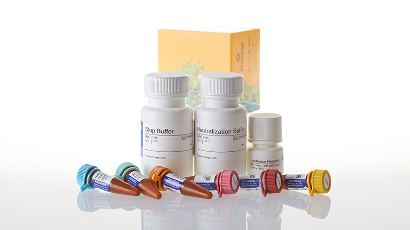
What's in the box?
| Item | Part # | Size |
|---|---|---|
Luciferase Reagent |
J151A | 1 × 5ml |
Stop Buffer |
J152A | 1 × 15ml |
Neutralization Buffer |
J153A | 1 × 15ml |
NADP+ |
J154A | 1 × 50μl |
G6PDH |
J155A | 1 × 125μl |
2DG |
J156A | 1 × 250μl |
2DG6P Standard |
J157A | 1 × 50μl |
Reductase |
G884C | 1 × 25μl |
Reductase Substrate |
G885A | 1 × 55μl |
SDS
Search for SDSCertificate of Analysis
Use Restrictions
For Research Use Only. Not for Use in Diagnostic Procedures.Storage Conditions
U.S. Pat. Nos. 9,273,343 and 9,951,372, European Pat. No. 2751089, Japanese Pat. No. 6067019 and other patents pending.
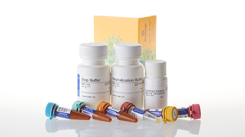
What's in the box?
| Item | Part # | Size | Concentration |
|---|---|---|---|
Luciferase Reagent |
J151B | 1 × 10ml | |
Stop Buffer |
J152A | 1 × 15ml | |
Neutralization Buffer |
J153A | 1 × 15ml | |
NADP+ |
J154B | 1 × 100μl | |
G6PDH |
J155B | 1 × 250μl | |
2DG |
J156A | 1 × 250μl | |
2DG6P Standard |
J157A | 1 × 50μl | |
Reductase |
G884A | 1 × 55μl | 6mg/ml |
Reductase Substrate |
G885A | 1 × 55μl |
SDS
Search for SDSCertificate of Analysis
Use Restrictions
For Research Use Only. Not for Use in Diagnostic Procedures.Storage Conditions
U.S. Pat. Nos. 9,273,343 and 9,951,372, European Pat. No. 2751089, Japanese Pat. No. 6067019 and other patents pending.
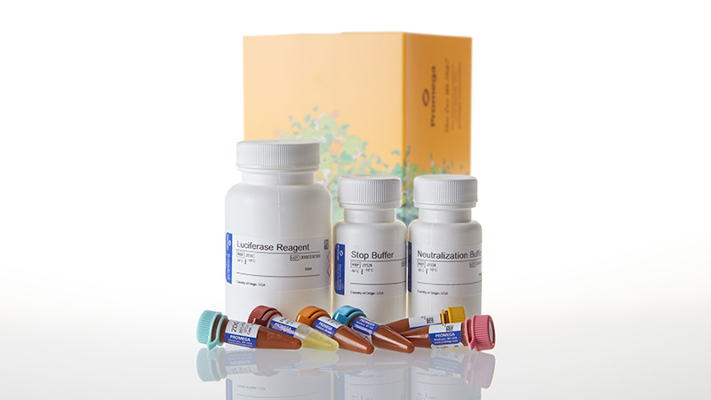
What's in the box?
| Item | Part # | Size | Concentration |
|---|---|---|---|
Luciferase Reagent |
J151C | 1 × 50ml | |
Stop Buffer |
J152A | 1 × 15ml | |
Neutralization Buffer |
J153A | 1 × 15ml | |
NADP+ |
J154C | 1 × 500μl | |
G6PDH |
J155C | 1 × 1.25ml | |
2DG |
J156A | 1 × 250μl | |
2DG6P Standard |
J157A | 1 × 50μl | |
Reductase |
G884B | 1 × 275μl | 6mg/ml |
Reductase Substrate |
G885A | 1 × 55μl |
SDS
Search for SDSCertificate of Analysis
Use Restrictions
For Research Use Only. Not for Use in Diagnostic Procedures.Storage Conditions
U.S. Pat. Nos. 9,273,343 and 9,951,372, European Pat. No. 2751089, Japanese Pat. No. 6067019 and other patents pending.
Related Products
Similar Products
Lactate-Glo™ Assay
Biolomunescent assay that quickly detect lactate from a variety of sample types.
J5021, J5022
Triglyceride-Glo™ Assay
Detects triglyceride levels by measuring glycerol that is released from an enzymatic reaction with a lipase.
J3160, J3161
Lumit® Glucagon Immunoassay
Quantitatively measures glucagon from cell culture or islet secretion samples using a fast, easy no-wash protocol.
W8020, W8022
Lumit® Insulin Immunoassay
Quantitatively measures insulin from cell culture or islet secretion samples using a fast, easy no-wash protocol.
W8010, W8012
Frequently Used With
GloMax® Discover System
High-performance microplate reader for detecting luminescence, fluorescence and absorbance.
GM3000
ROS-Glo™ H2O2 Assay
Sensitive, bioluminescent assay that measures the level of H2O2 in cell cultures.
G8820, G8821
GSH/GSSG-Glo™ Assay
Homogeneous assay to quantify total glutathione and glutathione ratios.
V6611, V6612
Mitochondrial ToxGlo™ Assay
An effective assay for predicting potential mitochondrial dysfunction.
G8000, G8001