CellTiter-Glo® 2.0 Cell Viability Assay
A Luminescent Cell Viability Assay for Fast, Easy, Everyday Use
- Quantifies viable cells in culture by measuring ATP—the universal biomarker of metabolically active cells
- Single, ready-to-use reagent—no substrate reconstitution required, reducing preparation time and variability
- Improved stability over the original CellTiter-Glo® assay: stable for repeated use over multiple days at 4°C
- Glow-type signal with >5-hour half-life enables flexible batch and automated plate processing
- Equivalent sensitivity and dynamic range to the original assay: detects as few as ~15–100 cells/well across 5 logs
- Optimized for high-throughput screening (HTS), automation and multiplex workflows
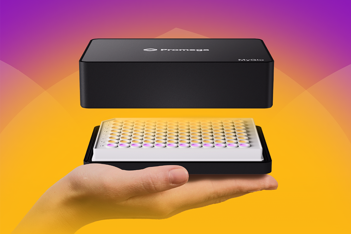
- Compatible with 96-, 384- and 1536-well formats; integrates with MyGlo® Reagent Reader


Catalog Number:
Choose a product
Size
Catalog Number: G9241
Catalog Number: G9242
Catalog Number: G9243
Catalog Number: MG1010

What Is the CellTiter-Glo® 2.0 Assay and How Does It Work?
The CellTiter-Glo® 2.0 Assay is a homogeneous, luminescent cell viability assay that determines the number of viable cells in culture by quantifying adenosine triphosphate (ATP)—the primary energy currency of living cells and a direct indicator of metabolic activity. It uses the same proven luciferase-based bioluminescent chemistry as the original CellTiter-Glo® Assay, but is reformulated as a single ready-to-use reagent for improved convenience, stability and throughput.
When CellTiter-Glo® 2.0 Reagent is added to cells, it simultaneously lyses the cells and drives a luciferase-luciferin reaction that produces a glow-type luminescent signal. The luminescence is directly proportional to the amount of ATP present, which in turn reflects the number of viable cells. Results are generated in approximately 10 minutes with a single reagent addition.
The CellTiter-Glo® 2.0 Assay determines the number of viable cells in culture by quantifying ATP, which indicates the presence of metabolically active cells. Luminescence readout is directly proportional to the number of viable cells in culture.

Why Was the CellTiter-Glo® 2.0 Assay Developed?
The original CellTiter-Glo® Assay requires reconstituting a lyophilized substrate in a separate buffer before use, limiting how long the reconstituted reagent can be stored. The CellTiter-Glo® 2.0 Assay was reformulated to address this limitation. The ready-to-use format eliminates the reconstitution step, maintains reagent stability over multiple days at 4°C and simplifies workflows for laboratories running repeated or large-scale screening campaigns.
What Are the Main Differences between the CellTiter-Glo® 2.0 Assay and the Original Version?
The CellTiter-Glo® 2.0 Assay delivers the same sensitivity, dynamic range, and luminescent signal as the original, with three meaningful operational improvements: a simplified single-reagent format, extended reagent stability and better suitability for multi-day high-throughput workflows.
| Feature | CellTiter-Glo® (Original) Assay | CellTiter-Glo® 2.0 Assay |
|---|---|---|
| Reagent format | Two-component: lyophilized substrate + buffer (requires reconstitution) | Single, ready-to-use solution (no reconstitution required) |
| Preparation time | ~30min (substrate reconstitution + equilibration) | ~15min (equilibration only) |
| Reagent stability (4°C) | Reconstituted: 48h (~5% loss); 4d (~20% loss) | Stable for repeated use over multiple days at 4°C |
| Sensitivity | Detects as few as 15 cells/well (384-well format) | Detects as few as 15 cells/well (384-well format) |
| Linear dynamic range | Up to 5 logs | Up to 5 logs |
| Signal type | Glow-type; >5h half-life | Glow-type; >5h half-life |
| HTS/automation | Yes: compatible with 96/384/1536-well formats | Yes: optimized for repeated batch use and automation |
| Multiplex compatibility | Standard | Improved; recommended for multiplex workflows |
| Best use case | Standard single-run viability and cytotoxicity assays | High-throughput, multi-day screening campaigns, automation |
Is Data Reproducibility Improved in the CellTiter-Glo® 2.0 Assay?
Data quality—sensitivity, linearity and signal stability—is equivalent between versions. Reproducibility improvements in the CellTiter-Glo® 2.0 Assay are primarily operational. By eliminating the substrate reconstitution step, the assay removes a source of plate-to-plate variability introduced when different users prepare reagent batches at different times. The stable, ready-to-use format also ensures that the reagent used on day 1 and day 5 of a screening campaign performs identically.
Are There Differences in Reagent Compatibility between Versions?
CellTiter-Glo® and CellTiter-Glo® 2.0 Assays should not be mixed. The two formulations contain different stabilizer systems, and mixing may produce unpredictable signal characteristics. If transitioning from the original to 2.0 within a validated assay, a side-by-side bridging experiment is recommended to confirm equivalence in your specific cell model and assay format before fully switching.
Does the CellTiter-Glo® 2.0 Assay Offer Faster Results than the Original?
Total assay time from reagent addition to luminescence measurement is equivalent (approximately 10–12 minutes). The speed improvement with the CellTiter-Glo® 2.0 Assay is in preparation time: eliminating reconstitution reduces setup by approximately 15–30 minutes per session, which accumulates meaningfully over multi-day screening campaigns.
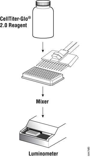
Simple Assay Setup and Enhanced Stability
A single, ready-to-use reagent and simple "add-mix-measure" protocol mean there's no need to mix components.
The CellTiter-Glo® 2.0 reagent is stable at room temperature and at 4°C, allowing fast and easy assay implementation, and eliminating the thawing and mixing required with other ATP assays. Enhanced stability also allows convenient storage.
| Percent Activity Remaining | CellTiter-Glo® 2.0 Reagent Stability | CellTiter-Glo® "Original" Reagent Stability |
|---|---|---|
| >85% Activity at 22°C | 7 days | 12 hours |
| >85% Activity at 4°C | 2 months | 3.5 days |
Assay Performance
The CellTiter-Glo® 2.0 Cell Viability Assay exhibits the same high performance as the classic CellTiter-Glo® Assay, making it suitable for use from benchtop scale to high-throughput screening.


High sensitivity and broad linearity. Luminescence measured with the CellTiter-Glo® 2.0 Assay is proportional to the number of viable Jurkat cells in culture over three orders of magnitude in 96-well, 384-well and 1536-well plates.

High sensitivity of the assay system enables reliable performance even from low cell densities. Jurkat cells were seeded in all-white 96-well plates at 400, 2,000 and 10,000 cells/well and were treated with Bortezomib for 20 hours. Viability was measured with CellTiter-Glo® 2.0 and MyGlo® Reagent Reader
Multiplex With Other Cell Viability or Cytotoxicity Assays
CellTiter-Glo® 2.0 cell viability assay can be multiplexed with other assays, reducing cell culture costs and labor. Multiplexing assays in the same sample well allows complementary measures of cell health to correct for errors in culturing, plating or assay chemistry interferences.
In the example shown here, CellTox™ Green cytotoxicity assay reagent was added to bortezomib-treated K562 cells after 48 hours of exposure. Fluorescence associated with cytotoxicity was measured. The CellTiter-Glo® 2.0 assay reagent was added and luminescence measured as an indicator of cell proliferation.

Case Study
Automated Assay Workflow for Mitochondrial Dysfunction
Challenge:
Louise Young, a research fellow at the University of Strathclyde, studies mitochondria’s role in Alzheimer’s and cancer. Her team needed a reliable, reproducible method to measure ROS output while screening compounds to decrease ROS levels. Traditional assays had wash steps, causing variability.
Result:
Using ROS-Glo™ H2O2 Assay, the team achieved faster turnaround with a no-wash protocol, greater reproducibility through luminescent readout and improved efficiency for quicker data collection.
Their workflow also includes RealTime-Glo™ Annexin V Assay for apoptosis/necrosis and CellTiter-Glo® 2.0 Assay for cell viability. Several compounds successfully reduced ROS, guiding future research into their protein targets.
Protocols
Frequently Asked Questions
What are the main differences between CellTiter-Glo® 2.0 and the original CellTiter-Glo® Assay?
The CellTiter-Glo® 2.0 Assay uses a single, ready-to-use liquid reagent that eliminates the substrate reconstitution step required with the original two-component kit. It also offers improved stability at 4°C over multiple days, making it better suited for repeated use across extended screening campaigns. Assay sensitivity, dynamic range, and signal half-life are equivalent between versions.
| Feature | CellTiter-Glo® (Original) Assay | CellTiter-Glo® 2.0 Assay |
|---|---|---|
| Reagent format | Two-component: lyophilized substrate + buffer (requires reconstitution) | Single, ready-to-use solution (no reconstitution required) |
| Preparation time | ~30min (substrate reconstitution + equilibration) | ~15min (equilibration only) |
| Reagent stability (4°C) | Reconstituted: 48h (~5% loss); 4d (~20% loss) | Stable for repeated use over multiple days at 4°C |
| Sensitivity | Detects as few as 15 cells/well (384-well format) | Detects as few as 15 cells/well (384-well format) |
| Linear dynamic range | Up to 5 logs | Up to 5 logs |
| Signal type | Glow-type; >5h half-life | Glow-type; >5h half-life |
| HTS/automation | Yes: compatible with 96/384/1536-well formats | Yes: optimized for repeated batch use and automation |
| Multiplex compatibility | Standard | Improved; recommended for multiplex workflows |
| Best use case | Standard single-run viability and cytotoxicity assays | High-throughput, multi-day screening campaigns, automation |
Is the sensitivity of the CellTiter-Glo® 2.0 Assay the same as the original?
Yes. The CellTiter-Glo® 2.0 Assay retains the same high sensitivity as the original assay—capable of detecting as few as approximately 15–100 cells/well depending on plate format—with a linear dynamic range spanning up to 5 logs. The reformulation focused on improving stability and workflow, not changing core assay chemistry.
How stable is the CellTiter-Glo® 2.0 Assay compared to the original?
The CellTiter-Glo® 2.0 Assay is stable for repeated use over multiple days when stored at 4°C after opening. The original CellTiter-Glo® reagent, once reconstituted, is stable for 48 hours at 4°C with approximately 5% signal loss, or 4 days with approximately 20% signal loss—requiring preparation close to the time of use.
Which version is recommended for high-throughput screening?
The CellTiter-Glo® 2.0 Assay is the recommended version for high-throughput screening and automated workflows. The ready-to-use format is more compatible with robotic liquid handlers, reduces batch-to-batch preparation variability, and maintains consistent performance across multi-day plate runs without requiring fresh reagent preparation each session.
Can the CellTiter-Glo® 2.0 Assay be used with 3D cell cultures?
The CellTiter-Glo® 2.0 Assay is not optimized for 3D spheroid or organoid models. For 3D cultures, Promega recommends CellTiter-Glo® 3D Cell Viability Assay, which uses an enhanced lysis buffer specifically formulated to penetrate 3D tumor spheroid structures and release ATP from cells throughout the spheroid.
What is the best cell viability assay for high-throughput drug screening?
ATP-based luminescence assays, such as the CellTiter-Glo® 2.0 Assay, are the preferred platform for HTS drug screening because they offer the highest sensitivity (detecting 15–100 cells/well), the simplest protocol (single reagent addition, 10-minute read time), and the lowest coefficient of variation across plate formats. Colorimetric assays (MTT, MTS) require additional processing steps and offer lower sensitivity at miniaturized volumes.
Are there cost differences between CellTiter-Glo® 2.0 and the original?
List pricing for CellTiter-Glo® 2.0 is modestly higher than the original per volume, reflecting the improved formulation and ready-to-use format. However, for high-throughput laboratories, the time savings from eliminated reconstitution steps and the ability to use reagent across multiple days may offset the per-assay cost difference. Contact your Promega representative for volume pricing options.
What should I do if CellTiter-Glo® 2.0 signals are inconsistent between plates?
Inconsistent signals across plates are most commonly caused by temperature differences between the plate and reagent at the time of addition (ensure both are equilibrated to room temperature for 30 minutes before mixing), insufficient cell lysis (increase mixing time or speed), or signal read outside the stable window (read plates within 10 minutes to 1 hour after stabilization). Confirm that white-walled plates are in use, as plate color significantly impacts luminescence signal intensity.
Specifications
Catalog Number:
What's in the box?
| Item | Part # | Size |
|---|---|---|
CellTiter-Glo® 2.0 Reagent |
G924A | 1 × 10ml |
SDS
Search for SDSCertificate of Analysis
Use Restrictions
For Research Use Only. Not for Use in Diagnostic Procedures.Storage Conditions
What's in the box?
| Item | Part # | Size |
|---|---|---|
CellTiter-Glo® 2.0 Reagent |
G924B | 1 × 100ml |
SDS
Search for SDSCertificate of Analysis
Use Restrictions
For Research Use Only. Not for Use in Diagnostic Procedures.Storage Conditions
What's in the box?
| Item | Part # | Size |
|---|---|---|
CellTiter-Glo® 2.0 Reagent |
G924C | 1 × 500ml |
SDS
Search for SDSCertificate of Analysis
Use Restrictions
For Research Use Only. Not for Use in Diagnostic Procedures.Storage Conditions
What's in the box?
| Item | Part # | Size | Available Separately |
|---|---|---|---|
MyGlo™ Reagent Reader
|
MG1000 | 1 × 1 each | |
CellTiter-Glo® 2.0 Reagent |
G9241 | 1 × 10ml | View Product |
SDS
Search for SDSCertificate of Analysis
Use Restrictions
For Research Use Only. Not for Use in Diagnostic Procedures.Storage Conditions
See Protocol for detailed storage recommendations.Resources
Articles
Citations
-
Effective photo-enhancement of cellular activity of fluorophore-octaarginine antisense PNA conjugates correlates with singlet oxygen formation, endosomal escape and chromophore lipophilicity.
2018
Sci. Rep.
-- -
Glutamine drives glutathione synthesis and contributes to radiation sensitivity of A549 and H460 lung cancer cell lines.
2016
Biochim. Biophys. Acta
-- -
DUX4-induced dsRNA and MYC mRNA stabilization activate apoptotic pathways in human cell models of facioscapulohumeral dystrophy.
2018
PLoS Genet.
-- -
Glucose starvation induces LKB1-AMPK-mediated MMP-9 expression in cancer cells.
2018
Sci. Rep.
-- -
miR-195 targets cyclin D3 and survivin to modulate the tumorigenesis of non-small cell lung cancer.
2018
Cell Death Dis.
-- -
In vitro ischemia decreases histone H4K16 acetylation in neural cells.
2015
FEBS Lett.
-- -
Imaging oxygen in neural cell and tissue models by means of anionic cell-permeable phosphorescent nanoparticles.
2015
Cell Molec. Life Sci.
-- -
Development and validation of a luminescence-base, medium throughput assay for drug screening in Schistosoma mansoni.
2015
PLOS Negl. Trop. Dis.
-- -
NRF2 Regulates HER1 Signaling Pathway to Modulate the Sensitivity of Ovarian Cancer Cells to Lapatinib and Erlotinib.
2017
Oxid. Med. Cell Longev.
-- -
The cellular stress proteins CHCHD10 and MNRR1 (CHCHD2): Partners in mitochondrial and nuclear function and dysfunction
2018
J. Biol. Chem.
-- -
Sensitivity and engineered resistance of myeloid leukemia cells to BRD9 inhibition.
2016
Nat Chem. Biol.
-- -
Traditional and systems biology based drug discovery for the rare tumor syndrome neurofibromatosis type 2.
2018
PLos ONE
-- -
Gas6-Axl signaling in presence of Sunitinib is enhanced, diversified and sustained in renal tumor cells, resulting in tumor-progressive advantages.
2017
Exp. Cell Res.
-- - See all citations
Compare Products
|
Cell Viability (ATP) |
Cell Viability (ATP) |
3D Cell Viability (ATP) |
Real-Time Viability |
Non-Lytic Viability |
Cell Proliferation |
|
|---|---|---|---|---|---|---|
| Best Use | Routine viability & HTS screening | Established protocols using original formulation | 3D spheroids, organoids, microtissues | Kinetic monitoring over time; downstream multiplexing | Multiplex first step; cells needed for follow-up assays | True proliferation readout independent of metabolism |
| Key Decision Points | ||||||
| Measures | Viability | Viability | Viability (3D) | Viability (kinetic) | Viability | Proliferation |
| Cells alive after assay? | ✗ Lytic | ✗ Lytic | ✗ Lytic | ✓ Non-lytic | ✓ Non-lytic | ✗ Lytic |
| Multiplexing compatible? | LimitedLytic—must be terminal step | LimitedLytic—must be terminal step | LimitedLytic—must be terminal step | ✓ ExcellentNon-lytic; pair with any downstream assay | ✓ ExcellentNon-lytic; pair with Caspase-Glo®, CTG, etc. | ModerateCan multiplex with CellTox™ Green or other fluorescent readouts |
| Real-time monitoring? | ✗ Endpoint | ✗ Endpoint | ✗ Endpoint | ✓ Up to 72hRead same wells repeatedly | ✗ Endpoint | ✗ Endpoint |
| 3D culture compatible? | PartialWorks for small spheroids; use 3D version for dense structures | PartialSame as 2.0 | ✓ OptimizedEnhanced lysis for dense 3D structures | PartialSubstrate must penetrate; best for small/loose 3D models | PartialSubstrate access may be limited in dense 3D | ✓ YesDetects Ki-67 in cell lysates from any culture format |
| Assay Attributes | ||||||
| Assay Principle | ATP quantitation (luciferase/luciferin) | ATP quantitation (luciferase/luciferin) | ATP quantitation (enhanced lysis for 3D) | Metabolic reduction of pro-substrate to luciferase substrate | Live-cell protease activity (GF-AFC cleavage) | Ki-67 immunodetection via NanoBiT® complementation |
| Detection Mode | Luminescence | Luminescence | Luminescence | Luminescence | Fluorescence400Ex / 505Em | Luminescence |
| Reagent Format | Ready-to-use liquid | Buffer + lyophilized substrateRequires reconstitution | Ready-to-use liquid | 2 components(enzyme + substrate) | Single reagent | Antibody mix + detection reagent |
| Time to Result | 10min | 10min | ~30min | ContinuousFirst read: 1–2h after addition | 30min | ~2h |
| Practical Considerations | ||||||
| Plate Formats | 96, 384, 1536 | 96, 384, 1536 | 96, 384 | 96, 384 | 96, 384 | 96, 384 |
| HTS Suitability | ✓ Excellent1536-well capable; fast protocol | ✓ Excellent1536-well capable | ✓ Good | ModerateRequires kinetic reader scheduling | ✓ Good | ✓ Good |
| Sensitivity (96-well) | ~15 cells/well | ~10 cells/well | Spheroid-dependent | <100 cells/well | ~40 cells/well | Cell line-dependent |
Similar Products
CellTiter-Glo® 3D Cell Viability Assay
A homogeneous method optimized to assess viability in 3D cell culture.
G9681, G9682, G9683
RealTime-Glo™ MT Cell Viability Assay
A bioluminescent method to kinetically monitor viability in cell culture up to 72 hours.
G9711, G9712, G9713
CellTiter-Fluor™ Cell Viability Assay
A non-lytic, fluorescent cell viability assay with multiplex capability.
G6080, G6081, G6082
Lumit® hKi-67 Immunoassay for Cell Proliferation
Easy add-mix-measure plate-based assay for tracking Ki-67 without washing steps or specialized detection equipment.
CS3076A01
Frequently Used With
GloMax® Discover System
High-performance microplate reader for detecting luminescence, fluorescence and absorbance.
GM3000
CellTox™ Green Cytotoxicity Assay
Measures changes in membrane integrity. Kinetically monitors cytotoxicity up to 72 hours with multiplex capability.
G8741, G8742, G8743, G8731
RealTime-Glo™ Apoptosis and Necrosis Assay
A one-step, plate-based assay for continually monitoring apoptosis progression.
JA1011, JA1012, JA1000, JA1001
Caspase-Glo® 3/7 Assay System
An easy-to-use, plate-based luminescent assay for detecting caspase-3/7 activity.
G8090, G8091, G8093, G8092, MG1010
Product Citations
Recent publications that mention the use of this product.