
Biologically Relevant Models to Advance your Research
Discover Glo 2023 - Biologically Relevant Models to Advance your Research
Now Available On Demand
The Discover Glo Conference is a free-of-charge premier event for the international scientific community to share knowledge and advancements in bioluminescent discovery tools and approaches for small molecules and biologics.
Following a simultaneous in-person and virtual experience to delegates across Europe, we are please to bring you the recordings of this day.
With a diverse lineup of international keynote speakers, cutting-edge presentations, and networking opportunities, this conference held its promises of being an invaluable opportunity for scientists, researchers, and industry professionals alike.
So why not sit back in your office, lab, or home and view the excellent scientific presentations and exchanges?
Do you have any questions? You can ask them in the presentations and we will relay them to our speakers.
About Discover Glo 2023
This year's Discover Glo Conference is now available as on demand webinars, after a hybrid event.
Watch the sessions online from the comfort of your lab or office.
Research platforms continue to evolve allowing for new biological models that more closely mimic the natural context, while still providing the benefits of throughput and ease of use that come with traditional approaches. Work historically done in biochemical formats can now be done in 2D cell models, which are being expanded to include more relevant cell types such as primary and iPSCs. 3D cell culture, co-culture and organoids are also increasingly being used to create models that more closely mimic the in vivo environment, providing methods to bridge the gap between biochemical or 2D cell culture and the in vivo biology being studied. In addition, the rapid growth of endogenous genome engineering allows for the creation of disease-specific models or reporter knock-ins at endogenous loci that can be used in 2D or 3D formats. Finally, new tools are being created to allow for increased mechanistic studies directly in in vivo animal models, improving the clinical relevance of this research.
These advances provide scientists with new research options that more accurately reflect living systems, minimize perturbance to the biology being studied, and yield results that will be more readily translatable in the future. In this event we heard about current research using models along this continuum that demonstrate successful implementation of new tools to create increasingly relevant and predictive results.
Watch the Recorded Sessions
This year's Discover Glo Conference offers a full day of scientific talks delivered by renowned international speakers from across Promega and its customer network.
You can now view all the session from the confort of your lab, office or home. We have included an option to email us your questions. We'll pass them along to our speakers and get back to you with the answers as quickly as possible.
You do not need specific software installed, just followin the links below to access each sessions, or use the button below to register for multiple ones.
Agenda
Biologically Relevant Models to Advance your Research
| Order | Duration | Talk | Link |
|---|---|---|---|
| 1 | 4 minutes | Welcome & opening remarks Poncho Meisenheimer Ph.D. Vice President of R&D, Promega Corp. |
Click Here |
| 2 | 48 minutes |
Exploring Endogenous Cell Biology with CRISPR-Mediated Bioluminescent Tagging |
Click Here |
| 3 | 35 minutes | A transgenic mouse model for multi-scale visualization of stress activation Cédric Chaveroux, Ph.D. Research fellow CNRS, leader of the “Nutritional stress response” group, Cancer Research Center of Lyon Read Abstract | View Bio |
Click Here |
| 4 | 40 minutes | From Petri Dishes to Chips: How Biochemical Assays Enhance Organ-on-Chip Technology Flavio Bonanini Early Stage Researcher, MIMETAS Read Abstract | View Bio Coming soon |
Contact us for access |
| 5 | 25 minutes |
Integrating bioluminescence and microfluidic chips for single-cell imaging and multiscale analysis |
Click Here |
| 6 | 45 minutes | Studying compound binding kinetics using NanoBRET distinguishes between highly similar targets in cells Anne Jackson Ph.D. Mechanistic and Structural Biology, Discovery Sciences, R&D, AstraZeneca, Cambridge Read Abstract | View Bio Coming soon |
Click Here |
| 7 | 51 minutes |
Real-Time Cell Health Assays Reveal More Relevant Biology * |
Click Here |
Speakers
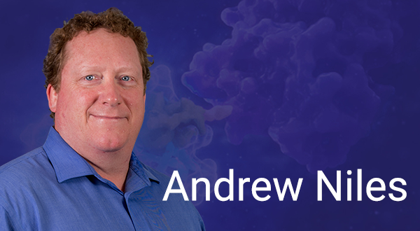
Andrew L. Niles
Senior Research ScientistAdvanced Technology Group
Promega Corp., Madison WI
USA
Exploring Endogenous Cell Biology with CRISPR-Mediated Bioluminescent Tagging
Stably integrated transgenes and tags have been useful for interrogating basic aspects of cell biology, but often fail to fully reflect physiologically relevant parameters relating to binding partners and interactors. These known limitations may adversely skew data set interpretation. CRISPR-mediated methods now allow for specific and precise incorporation of tags into native loci (“knockins”), thus offering notable and incremental improvement in achieving true endogenous biology. This seminar describes our efforts to improve gene editing efficiencies and workflows, means to identify and characterize those edits, and to demonstrate their utility using well-validated cancer pathway protein:protein interactions. Specific emphasis will be focused on our luciferase and luciferase complementation tagging systems and the relative merits or limitations of each approach.
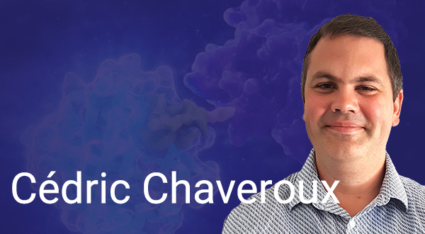
Cédric Chaveroux, Ph.D.
Research fellow CNRSLeader of the “Nutritional Stress Response” group, Dr. T . Renno’s team
Cancer Research Center of Lyon
France
A transgenic mouse model for multi-scale visualization of stress activation
Cells are continuously exposed to microenvironmental stressors. The ensuing adaptive response relies on various molecular sensors that activate signaling pathways governing transcriptional and translational programs. These programs then support cellular plasticity in the face of stress. The integrated stress response (ISR) axis represents a major signaling pathway controlling various cellular functions involved in cellular plasticity. However, the inducibility of ISR is heterogeneous within the organism and rather tissue and cell type specific. Thus, the study of how the different cells comprising the tissue of interest respond to stress requires models that can track the degree of activation of stress pathways at all scales of the organism. To address this issue, we have developed a transgenic mouse model that allows the visualization of ISR activity at the organism, tissue and cell level. To illustrate the interest of this technology based on bioluminescence acquisition, our results show how stem or differentiated epithelial cells differentially induce ISR following proteostasis stress in derived intestinal organoids. Overall, this transgenic model is an interesting resource to monitor and study the degree of stress activation and reduce the use of animals in preclinical studies.
Dr. Chaveroux completed his Ph.D. in Nutrition at Clermont University in 2009. He stayed on at McGill University as a postdoctoral fellow in the Giguere lab and then in the Fafournoux lab until joining the Cancer Research Center of Lyon in 2016. Cédric’s research is investigating the molecular mechanisms linking nutritional stresses and cellular plasticity. He is particularly focusing on the response to ribotoxic and ER stresses and the contribution of the associated signaling pathway to translational reprogramming and cell adaptation.
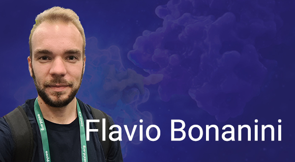
Flavio Bonanini
Early Stage Researcher
MIMETAS, Oegstgeest
The Netherlands
From Petri Dishes to Chips: How Biochemical Assays Enhance Organ-on-Chip Technology
The successful translation of pre-clinical research results into clinical practice remains a significant challenge for the pharmaceutical industry. Clinical trials often fail, have limited efficacy, or induce toxic side effects that were not anticipated. This is in part due to inadequate pre-clinical models, such as two-dimensional cell cultures, which do not mimic the complex environment cells find in vivo. Recent advances in organ-on-a-chip technology offer a promising avenue to model important aspects of human physiology in vitro. MIMETAS' OrganoPlate® is an organ-on-a-chip platform that allows for controlled and complex cultivation of cellular systems in three dimensions, mimicking architecture, mechanical properties, cellular composition and biochemical functionalities of healthy and diseased organs. To effectively harness the potential of this technology, sensitive, robust, reproducible, and interpretable assays are necessary for the study of complex tissue cultures. These assays must be able to accurately measure key physiological parameters and responses to treatments, allowing for the identification of potential drug candidates and the prediction of clinical outcomes. The integration of organ-on-a-chip technology and reliable assays could transform pre-clinical drug development by providing accurate and predictive models of human physiology.
I grew up in Southern Switzerland and relocated to Zurich in 2012 to commence my undergraduate studies in Biology at the Swiss Federal Institute of Technology (ETH). Subsequently, I pursued a Master's degree in Biomedical Engineering, with a specialization in tissue engineering, also at ETH, and successfully completed my studies in 2018.
From 2019, I have been a Marie Sklodowska-Curie fellow student affiliated with MIMETAS, a biotech company located in Leiden, Netherlands, as part of the PoLiMeR consortium. In my current role, I focus on liver models and contribute to the development of a throughput-capable organ-on-chip culture platform, which MIMETAS specializes in developing and commercializing.
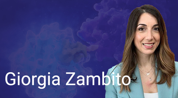
Giorgia Zambito Ph.D.
Scientist in Cancer Immunology, Optical Molecular Imaging and NanotechnologyErasmus Medical Center, Rotterdam
The Netherlands
Incorporating non-invasive biosensing features in organ-on-chip models is of paramount importance for a wider implementation of these advanced in vitro microfluidic platforms. Optical biosensors, based on Bioluminescence Imaging (BLI), enable continuous, non-invasive, and in-situ imaging of cells, tissues or miniaturized organs without the drawbacks of conventional fluorescence imaging. Here, we report the integration and optimization of BLI in organs-on-chips, for non-invasive imaging of multiple biological readouts. Indeed, we can detect Bioluminescence (BL) and fluorescence (FL) signals at single cell resolution on-chip post-addition of luciferin substrate under dynamic perfusion. The use of BLI with organ-on-chip technologies opens up the potential for simultaneous in situ detection with continuous monitoring of multicolor cell reporters. Moreover, this can be achieved in a non-invasive manner. BL has great promise as a highly desirable biosensor for studying organ on chip platforms.
Giorgia Zambito is a postdoc researcher with a strong background in pre-clinical research, with a specialization in optical molecular imaging.
In 2016, as an early-stage researcher, she deepened her knowledge of optical molecular imaging techniques and T-cell genetic engineering, when working at the department of Immunology at Leiden University Medical Center (LUMC)(NL).
In 2017, she started as a Ph.D. student developing MRI and optical imaging techniques for imaging immune cell localization in rodent models. Notably, she characterized a new luciferase mutant emitting a deep-red bioluminescence light, ideal to visualize living cells in deep organs and in real-time. She applied this technology to track multicolor cell populations as well as to study their behavior in a native environment. Altogether, these tools delineated the fate of immune cells targeting tumor metastases in a live organism, non-invasively.
Now, she is a postdoc researcher focusing her studies on microfluidic chip development that recapitulates in vitro, interactions between two joint tissues: cartilage and synovium. In this project, her expertise will help to delineate a toolbox of bioluminescent reporter genes used for real-time analysis of inflammatory and mechano-transduction read-outs.
Aside from the bench work, she is a committee member of the Dutch young Molecular Imaging Community (DyMIC), and she is involved in the Laboratory Efficiency Assessment Framework project (LEAF) to promote sustainable research in the lab.

Anne Jackson Ph.D.
Mechanistic and Structural Biology, Discovery Sciences, R&DAstraZeneca, Cambridge
United Kingdom
Studying compound binding kinetics using NanoBRET distinguishes between highly similar targets in cells
Compound selectivity between targets and off-targets is often usefully measured by comparing compound potency at equilibrium, but this does not always give the whole picture. When investigating the inhibition of two highly similar kinases, these measures at equilibrium showed little selectivity across a range of biochemical and cellular systems. When biophysical methods showed differences in binding kinetics, we investigated the compound behaviour in cells using NanoBRET target engagement assays in kinetic mode. These showed dramatic differences in rates of binding and the length of compound-target interaction between the two kinases, suggesting greater selectivity than previously suspected from the equilibrium data and informing the design of subsequent experiments to factor in the time-dependent nature of the compound activity. The impacts of these findings and learnings from generating and analysing large amounts of kinetic data will be discussed, along with the importance of understanding the assumptions of biochemical models when applying them to cellular settings.
